Fig. 2.

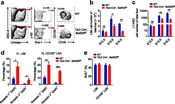
The expansion of FL HSCs in Tie2-Cre+, Baf200f/f embryos is severely impaired. a FACS profiles of LSK and LT-HSC (CD150+CD48−LSK) subsets in the FLs from E15.5 Tie2-Cre+, Baf200f/f embryos and WT littermates. b, c Absolute cell number of LSK cells (b) and LT-HSCs (c) from Tie2-Cre+, Baf200f/f embryos and WT littermates at indicated stages. d Percentage of apoptotic cells of LSK and CD150+ LSK cells from E14.5 Tie2-Cre+, Baf200f/f embryos (n = 4) and WT littermates (n = 5). The apoptosis was evaluated by using annexin V and DAPI. e Cell cycle status of LSK and CD150+ LSK cells from E14.5 Tie2-Cre+, Baf200f/f embryos (n = 4) and WT littermates (n = 5). No difference of BrdU+ cells was observed. Data are shown as means ± SEM. *P < 0.05; **P < 0.01; ***P < 0.001. ns indicates no significant difference
