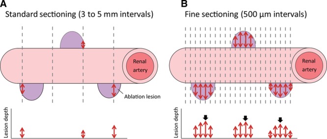
Figure 2.

Standard (A) and fine (B) sectioning methods for histological evaluation. A, With the standard method, because the histological section does not always capture the center of the ablation lesion (purple), the average measured lesion depth (red double arrows) was theoretically less than that of the true lesion depth. B, With the fine sectioning method, several sections were produced from 1 lesion, and the lesion depth was measured for all sections. The maximum value (black arrows) per lesion was used to calculate the averaged maximum lesion depth at each ablation setting.
