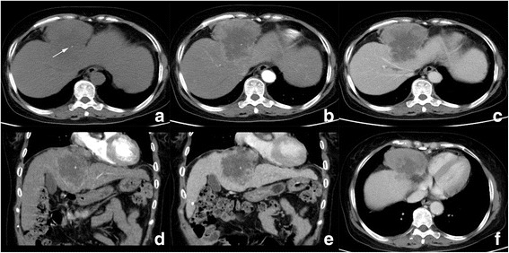
Fig. 4.

Computed tomography (CT) images of patient 2. a Unenhanced CT images reveal a hypodense mass in segment 4 with unclear margin and spot calcification (arrow). Tumor showed significant enhancement in peripheral portion in AP (b) and washout in PP (c); d and e are coronal reconstructed PP images, present large central part of cystic degeneration in the tumor; right phrenic angle lymphadenopathy is also observed (f, *)
