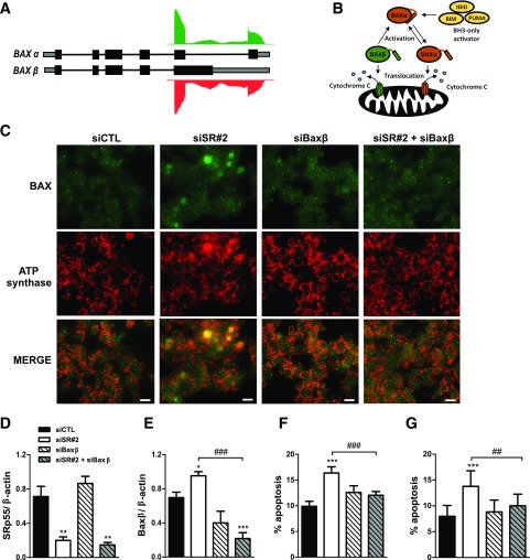
Figure 5.
SRp55 controls the expression of a constitutively active isoform of the apoptotic inducer BAX, contributing to increased β-cell apoptosis. A: Schematic representation of BAX isoforms α and β, and RNA sequencing reads in control and SRp55 KD cells mapping to the distal part of the gene. Boxes represent exons; gray represents untranslated regions; black represents coding regions; and solid lines represent introns. B: Model of activation of apoptosis by BAX α and BAX β isoforms proposed by Fu et al. (33). Upon apoptotic signaling, BH3-only molecules such as BIM activate BAX α to promote its translocation and oligomerization to the mitochondrial outer membrane, leading to cytochrome c release and apoptosis activation. On the other hand, BAX β spontaneously targets, oligomerizes, and permeabilizes mitochondria, behaving as a constitutively active isoform. C–G: Double KD of SRp55 and BAX β in EndoC-βH1 cells (C–F) and in human islets (G). Cells were transfected with siCTL, siSR#2, siBaxβ, or siSR#2 plus siBaxβ for 48 h. C: Fluorescence microscopy analysis of BAX and the mitochondrial marker ATP synthase in EndoC-βH1 cells, showing that SRp55 KD leads to increased translocation of BAX to the mitochondria, a phenomenon prevented by BAX β silencing. Scale bar = 1 µm. mRNA expression of SRp55 (D) and BAX β (E) was measured by quantitative RT-PCR and normalized by the housekeeping gene β-actin. mRNA expression values were normalized by the highest value of each experiment, considered as 1. Proportion of apoptotic cells in EndoC-βH1 cells (F) and in dispersed human islets (G). Results are the mean ± SEM of four or five independent experiments. *P < 0.05, **P < 0.01, and ***P < 0.001 vs. siCTL; ##P < 0.01 and ###P < 0.001 as indicated by bars (ANOVA followed by the Bonferroni post hoc test).

