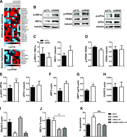
Figure 7.
SRp55 KD–induced ER stress contributes to β-cell death. A: Heat map showing AS and gene expression changes in genes involved in the ER-associated protein degradation process (top panel) and markers of the unfolded protein response (bottom panel). Red represents higher expression and blue, lower expression. B–E: Representative Western blotting (B) and densitometric measurements of total and phosphorylated forms of IRE1α (C), PERK (D), and eIF2α (E). mRNA expression of BIP (F), XBP1 spliced (G), and CHOP (H) after SRp55 KD was measured by quantitative RT-PCR (qRT-PCR) and normalized by the housekeeping gene β-actin. I–K: Double KD of SRp55 and IRE1α in EndoC-βH1 cells. Cells were transfected with siCTL, siSR#2, siIRE1α, or siSR#2 plus siIRE1α for 48 h. mRNA expression of SRp55 (I) and IRE1α (J) was measured by qRT-PCR and normalized by the housekeeping gene β-actin. mRNA expression values were normalized by the highest value of each experiment, considered as 1. K: The proportion of apoptotic cells was evaluated by Hoechst/propidium iodide staining. Results are the mean ± SEM of four to nine independent experiments. *P < 0.05, **P < 0.01, and ***P < 0.001 vs. siCTL; #P < 0.05 and ###P < 0.001 as indicated by bars (paired t test [C–H] or ANOVA followed by the Bonferroni post hoc test [I–K]).

