
Fig. 1.
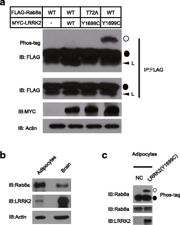
Phos-tag analysis of LRRK2 Y1699C mediated Rab8a phosphorylation. a 293 T cells were transfected with MYC-LRRK2 (Wild type or Y1699C), and FLAG-Rab8a (WT or T72A mutant). Anti-FLAG M2 beads were used for immunoprecipitation. The immunoprecipitated products were detected by using antibody against FLAG. Phosphorylation of overexpressed Rab8a was analysed by a Phos-tag assay (top panel). Equal levels of expression of FLAG-Rab8a and MYC-LRRK2 were confirmed by immunoblotting on normal gels using an anti-FLAG (second panel from the top) and anti-MYC (third panel from the top) antibodies respectively. Actin was used as a loading control (bottom panel). L represents light chain. ○ represents phosphorylated Rab8a. ● represents non-phosphorylated Rab8a. Similar results were obtained in at least two separate experiments. b The expression level of Rab8a and LRRK2 in adipocytes. c The phosphorylation of endogenous Rab8a in adipocytes. Differentiated adipocytes were transfected with LRRK2 (Y1699C)