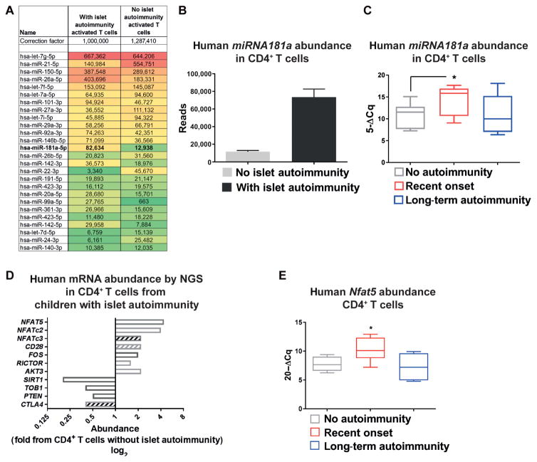
Fig. 2. miRNA181a targets NFAT5 in human CD4+ T cells.
(A) MicroRNA (miRNA) expression profiles in ex vivo CD4+ T cells with an activated phenotype from children with or without autoantibodies by next-generation sequencing (NGS) of pooled samples (n = 4 per group). A set of most abundant miRNAs relevant for T cell activation and or Treg induction is shown. (B) MiRNA181a reads by NGS as in (A) (n = 4 per group). (C) MiRNA181a abundance in ex vivo CD4+ T cells from children with different durations of autoimmunity (no autoimmunity, n = 9; recent onset of autoimmunity, n = 10; long-term autoimmunity, n = 5) by real-time quantitative polymerase chain reaction (RT-qPCR). (D) Abundance of signaling intermediates involved in T cell activation in ex vivo CD4+ T cells from autoantibody-negative or autoantibody-positive children by NGS from pooled samples (n = 4 per group). Open bars, predicted as direct targets of miRNA181a; hatched bars, not predicted as direct targets of miRNA181a. (E) Human Nfat5 mRNA abundance in ex vivo CD4+ T cells from individual children with or without islet autoimmunity by RT-qPCR (no autoimmunity, n = 6; recent onset of autoimmunity, n = 7; long-term autoimmunity, n = 6). Data are means ± SEM (B) or are presented as box and whisker plots with minimum to maximum values for data distribution (C and E). *P < 0.05, Student’s t test. PTEN, phosphatase and tensin homolog; NFAT5, nuclear factor of activated T cells 5; CTLA4, cytotoxic T lymphocyte–associated protein 4; TOB1, transducer of ERRB2 1.

