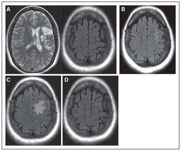
Figure 2.
A–D, MR brain scans from a 39-year-old female patient, transplanted with SB623 cells 2 years after a left middle cerebral artery stroke. (A left) Axial T2 FSE pretransplant showing the subcortical and cortical infarct. (A right) Pretransplant at higher axial level. (B) Day 1 post-transplant at higher axial level demonstrating small amount of blood in left frontal sulci. (C) Day 7 post-transplant at higher axial level showing new T2 FLAIR signal abnormality in left superior frontal gyrus adjacent to premotor gyrus. (D) Month 2 post-transplant at higher axial level showing resolution of T2 FLAIR signal abnormality. FLAIR indicates fluid-attenuated inversion recovery; FSE, fast spin echo; and MR, magnetic resonance.

