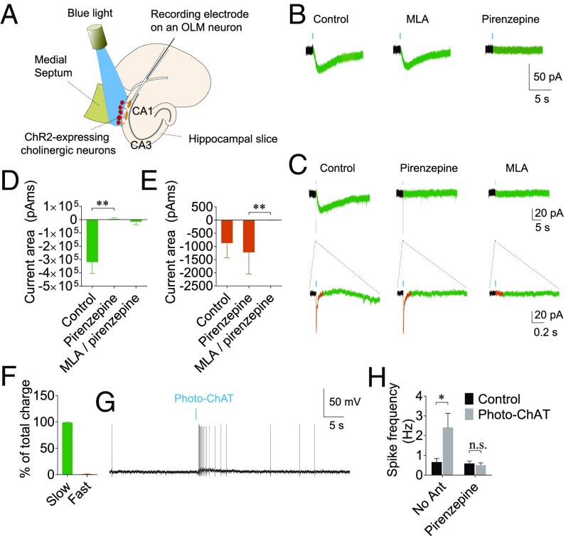
Fig. 2.
Cholinergic activation of OLM interneurons. (A) A schematic showing recording of AChR currents in OLM interneurons triggered by photostimulation of cholinergic neurons. (B) Photostimulation of cholinergic neurons (shown as blue bars, 0.27–2.7 mW LED on cholinergic cell bodies through the 4× objective or 60–600 µW LED light onto the cholinergic fibers through the submerged 40× objective) for 1–10 ms evoked slow inward currents (green) in the majority of OLM interneurons (∼93%). (C) In a subset of OLM cells recorded (∼60%), ACh also evoked fast inward currents (orange), which were sensitive to the α7 nAChR antagonist MLA but not to pirenzepine. (D) Average areas of slow currents evoked by photostimulation of cholinergic neurons, showing that the slow currents are mediated by the M1 muscarinic AChRs. (E) Average areas of fast currents evoked by photostimulation of cholinergic neurons showing that the fast currents are mediated by α7 nAChR and are blocked by MLA. (F) Summary bar graph showing the contributions of fast and slow components to the total charge. (G) A representative current-clamp recording trace showing that photostimulation of ChAT neurons caused an increase in spike frequency in OLM neurons. (H) Summary bar graph showing an increase in spike frequency induced by cholinergic neurons, which was abolished in the presence of pirenzepine. *P < 0.05; **P < 0.01.

