Fig. 5.
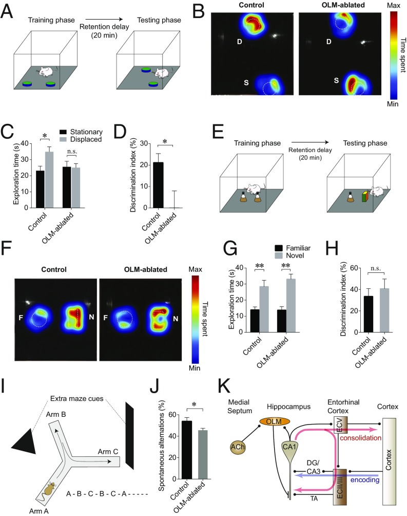
Ablation of OLM interneurons impairs the encoding of spatial memory. (A) A schematic drawing showing the OLT. During the training phase, a mouse was placed in an arena that contained two identical objects. After a 20-min retention delay, during which one of the objects was moved to a new location, the mouse was replaced in the arena, and the mouse’s exploration of the objects was recorded. (B) Heat maps showing that an OLM-ablated mouse, unlike the control mouse, does not show a preference for the object with a novel location (D, displaced) over the object that has not been moved (S, stationary). (C) Summary of exploration time for each object showing that the littermate controls spent significantly more time on the object with a novel location, whereas OLM-ablated mice did not show a preference for the object with a novel location. (D) The DI of OLM-ablated mice was significantly lower than that of control mice. (E) A schematic drawing showing the NORT. In this task one of the initial objects placed during the training phase was replaced with a novel object before the testing phase. (F) Heat maps showing that both control and OLM-ablated mice preferred the novel (N) over the familiar (F) object. (G) OLM-ablated mice spent significantly more time on exploring the novel object, as did the control mice. (H) Summary bar graph showing that DI of the OLM-ablated mice was not significantly different from that of controls. (I) OLM-ablated mice were tested for the spontaneous alternation task on a Y-maze with extramaze spatial cues to determine their spatial working memory. (J) OLM-ablated mice showed significantly reduced spontaneous alternation performance compared with their littermate controls. See also Figs. S4–S6. (K) A proposed model of cholinergic regulation of the hippocampus–EC circuit. ACh stimulates OLM interneurons in the hippocampus by activating M1 muscarinic AChRs expressed on OLM interneurons. The activation of OLM interneurons causes an increase in GABAergic synaptic inputs to the dendrites of CA1 pyramidal neurons in the stratum lacunosum moleculare layer, which effectively suppresses the temporoammonic (TA) pathway-induced firing of CA1 pyramidal neurons. This suppresses the memory-consolidation circuits (by decreasing output to the ECV), which then may allow CA1 pyramidal neurons to be available for regulation via the memory-encoding pathway. *P < 0.05; **P < 0.01.

