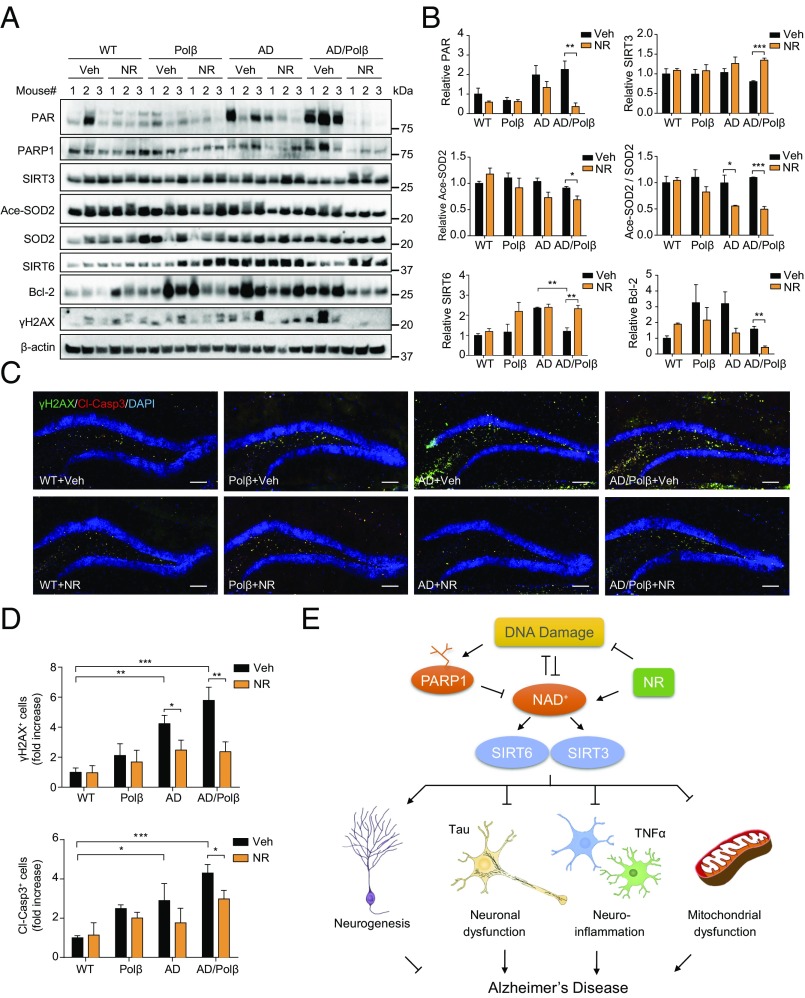
Fig. 5.
DNA damage and apoptosis are decreased after NR treatment. (A) Representative immunoblots of the indicated proteins from the hippocampus of WT, Polβ, AD, and AD/Polβ mice after treatment with vehicle or NR. Quantifications of data are shown in B and Fig. S7. (B) Quantification of immunoblots from the indicated proteins in A. (C) Representative images of γH2AX (green), cleaved-caspase 3 (red), and DAPI (blue) staining of DG sections from WT, Polβ, AD, and AD/Polβ mice treated with vehicle or NR. (Scale bars, 100 µm.) (D) Quantification of γH2AX+ cells and cleaved-caspase3+ cells from sections as in C. n = 5 mice per group. (E) Summary figures showing our proposed mechanisms of the relation among NAD+, DNA damage, sirtuin, and AD. For B and D, data are shown as mean ± SEM. *P < 0.05, **P < 0.01, ***P < 0.001.

