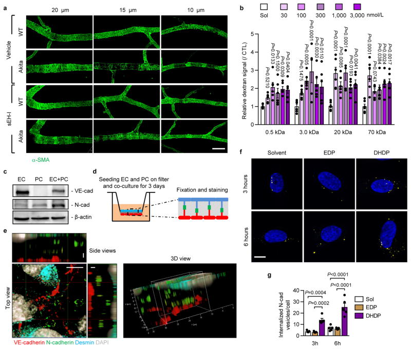
Extended Data Figure 4. Mural cell coverage and N-cadherin localization in endothelial cell-pericyte co-cultures.
a, Smooth muscle actin (α-SMA) showing mural cell coverage of 20 μm, 15 μm and 10 μm diameter retinal vessels; bar = 50 mm. Comparable results were obtained in retinas from 5 additional animals in each group. b, Permeability of human endothelial cells to dextrans of different molecular mass; n = 6 independent cell preparations. c, VE-cadherin and N-cadherin expression in endothelial cells (EC), pericytes (PC) or endothelial-pericyte co-cultures (EC+PC). Similar results were obtained in 3 different cell batches. For gel source data, see Supplementary Fig. 1. d, Cartoon showing the filter-based co-culture system studied. e, VE-cadherin (red), N-cadherin (green), desmin (cyan) and DAPI (grey) on transwell membranes with endothelial-pericyte co-cultures; bar = 5 μm in top view and 2 μm in side views. Similar observations were made in 2 additional experiments. f–g, Internalized N-cadherin (yellow) in pericytes treated with solvent, 19,20-EDP or 19,20-DHDP in the presence of sEH inhibitor; n = 5 independent experiments; bar = 10 μm. Data expressed as mean ± s.e.m. P values determined by 1way ANOVA.

