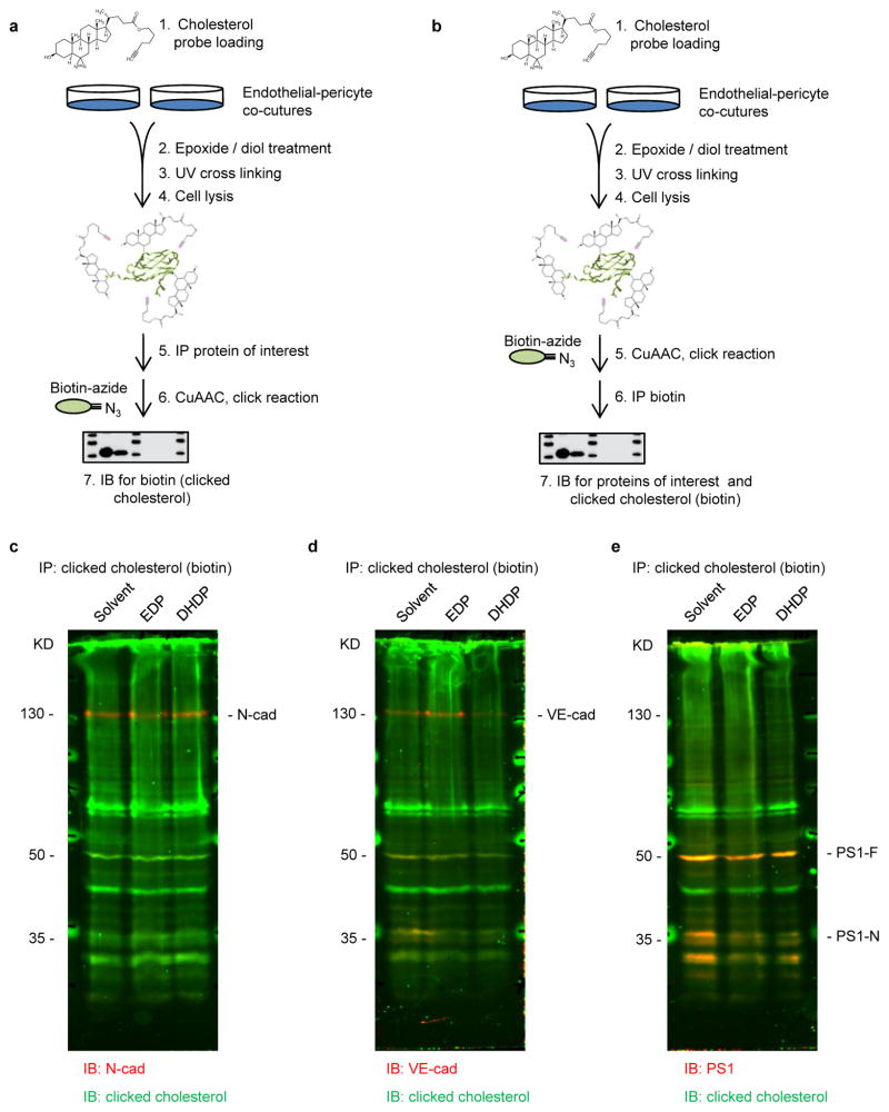
Extended Data Figure 5. Association of cholesterol with cadherins and PS1.
a, Experimental pipeline for studying the association of proteins with cholesterol in Fig. 3n–o. b, Reverse immunoprecipitation for studying the association of proteins with cholesterol. Note the difference in step 5 and 6 from experimental procedure in a. c–e, Representative Li-cor system images. Proteins clicked with cholesterol were visualized in green; i.e. (c) N-cadherin, (d) VE-cadherin and (e) presenilin1 (PS1-F: full length; PS1-N: N-terminal fragment) were detected on the same membrane and visualized in red. Comparable results were observed in 3 additional experiments.

