
Figure 4. Statistical analysis of PV+ neurons imaged using confocal microscopy.
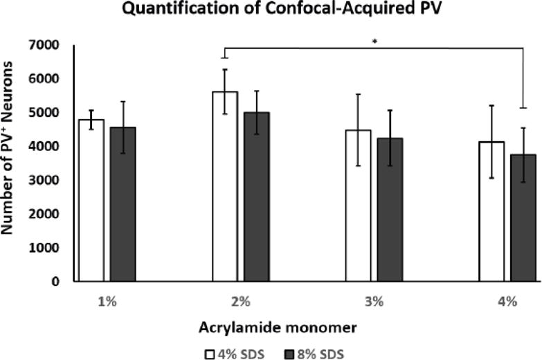
2% monomer/4% SDS showed a significantly higher number of detected PV+ neurons in vs. 4% monomer/8% SDS infused tissue (*p<0.02; 1M4S, n=4; all other groups, n=5, +/- SD). Abbreviations: PV; parvalbumin, SDS; sodium dodecyl sulfate.