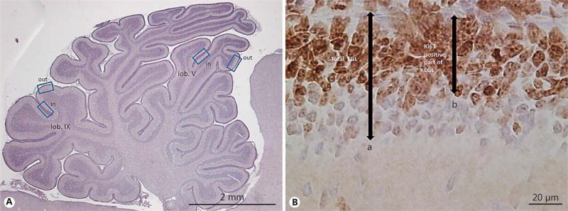
Fig. 1.
Measurement of external granular layer (EGL) width. A Cerebellar overview illustrating areas (outward and inward regions of lobuli V and IX) used for quantitative measurement of total and proliferative EGL width. B HE- and Ki67-stained section of the EGL illustrating measurements obtained for quantitative EGL analysis: (a) total EGL = width of proliferative EGL + inner zone of differentiated granular precursor cell (GPC) layer, and (b) proliferative EGL = width of Ki67-positive proliferating GPC layer.

