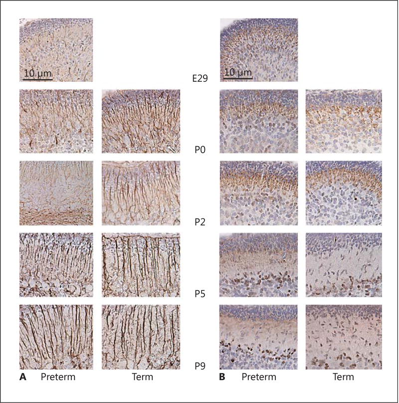
Fig. 5.
Cerebellar Bergman glia development and expression of cleaved caspase-3 in preterm (PT) and term (T) rabbit pups. A Immunostaining with GFAP was used to evaluate Bergman glia development in PT and T rabbit pups at each postnatal time point as described in Material and Methods. No clear differences in Bergman glia morphology were observed between the PT and T groups. At E29, GFAP-positive glial fibers were poorly defined and very scarce. With increasing postnatal age, glial fibers become more developed and confined to the molecular layer. B Staining for cleaved caspase-3 was applied to evaluate possible apoptosis during cerebellar development in the PT and T groups, as described in Material and Methods. Positive staining for cleaved caspase-3 (brown) was restricted to the radial fibers of the Bergman glia at E29, P0, and P2, and was predominantly located at the interface between the inner external granular layer and the molecular layer. At P5 and P9, staining for cleaved caspase-3 was restricted to the somata of cells, with a localization suggestive of Bergmann glia. The staining pattern for cleaved caspase-3 suggested a constitutive expression during Bergmann glia development and did not differ between the PT and T groups. Scale bar, 10 µm.

