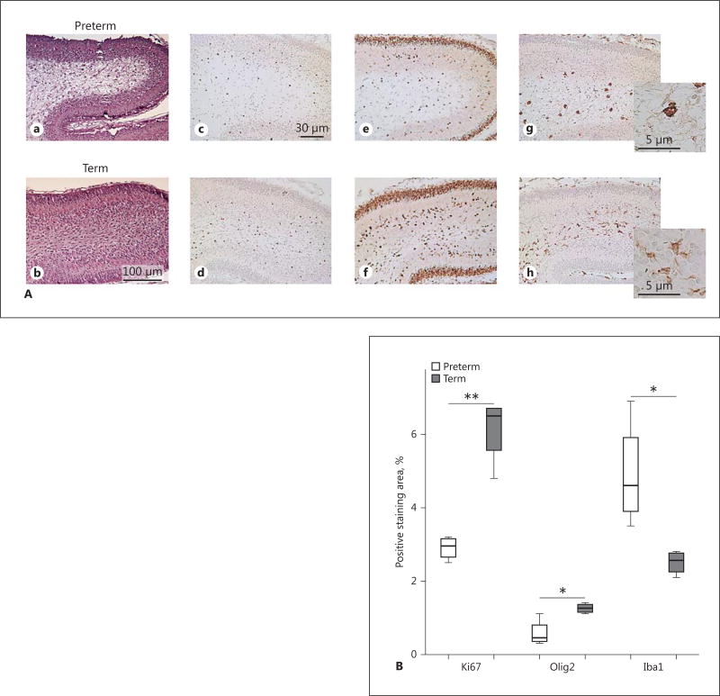
Fig. 6.
Cerebellar white-matter damage in preterm (PT) rabbit pups. A Cerebellar sections exhibited signs of cellular damage in cerebellar white matter in PT pups at P2 and P5. HE. Cerebellar white-matter damage was not observed in T pups. Immunostaining with antibodies against Olig2, Ki-67, and Iba1, respectively, were performed as described in Material and Methods to characterize proliferation and oligodendroglial and microglial cellular response in cerebellar white matter in PT and T rabbit pups. Upper row: HE (a), Olig2 (c), Ki67 (e) and Iba1 (g) staining, respectively, in a representative PT rabbit pup at P2. Signs of white-matter damage in the HE-stained section correspond to a marked reduction of proliferating cells, a reduced number of Olig2-positive cells, and an increased number of Iba1-positive microglia in the cerebellar white matter. Lower row: the corresponding immunostainings in a T pup at P2 (b, d, f, h) with no signs of white-matter damage for comparison. Scale bars: 100 µm (HE); 30 µm (Ki67, Olig2, and Iba1). Insets An activated ameboid Iba1-positive microglial morphology in the PT pup (g) compared to a quiescent ramified morphology in the T pup (h). B Quantitative analysis showed a decrease in staining for Olig2 (** p = 0.009) and Ki67 (* p = 0.03) and an increase in staining for Iba1 (* p = 0.04).

