Figure 4.

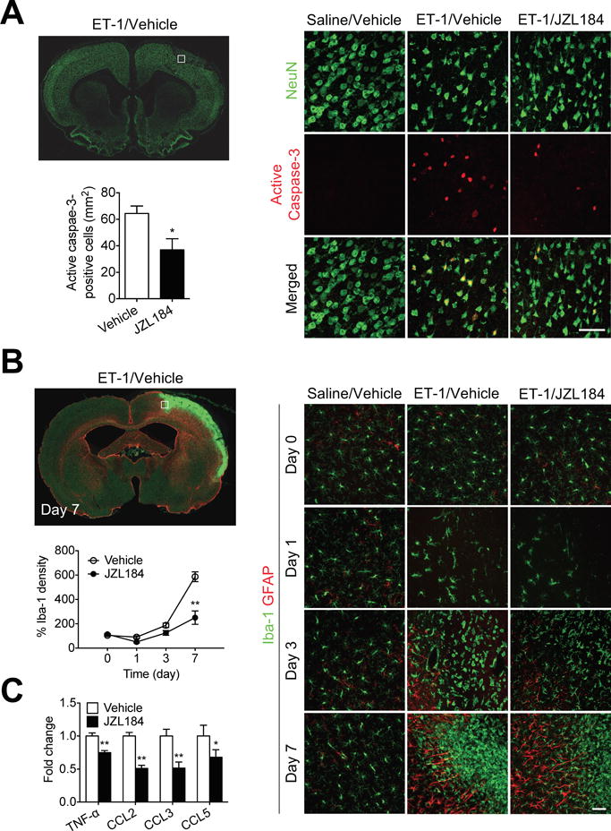
MAGL inhibitor JZL184 reduces neuronal injury and inflammation after stroke. A, Double-immunofluorescence staining of active caspase-3 (red) and neurons (NeuN, green) in the ipsilateral hemisphere of SHR at 1 d after ET-1-induced ischemic stroke. High-magnification images showing representative immunoreactivity in the cortex. Scale bar, 50 μm. Quantification of active caspase-3+ neurons in the ischemic hemisphere of vehicle- and JZL184-treated SHR. Data are expressed as mean ± SEM (n = 6/group). *P < 0.05 versus vehicle, unpaired t-test. B, Double-immunofluorescence staining of astrocytes (GFAP, red) and microglia (Iba-1, green) in the ipsilateral hemisphere of SHR at 1 d after ET-1-induced ischemic stroke. High-magnification images showing representative immunoreactivity in the cortex at 1, 3, and 7 d after ET-1-induced ischemic stroke. Scale bar, 50 μm. Quantification of the temporal density of Iba-1+ microglia in the peri-infarct regions of cortex at different time points. Data are expressed as mean ± SEM (n = 6/group). **P < 0.01 versus the respective vehicle group, two-way ANOVA followed by Bonferroni’s multiple comparison test. C, Relative protein expression of TNF-α, CCL2, CCL3, and CCL5 in the cortices of vehicle- and JZL184-treated SHR at 1 d after ET-1-induced stroke. Data are expressed as mean ± SEM (n = 6/group). *P < 0.05, **P < 0.01 versus vehicle, unpaired t-test.
