
Figure 4.
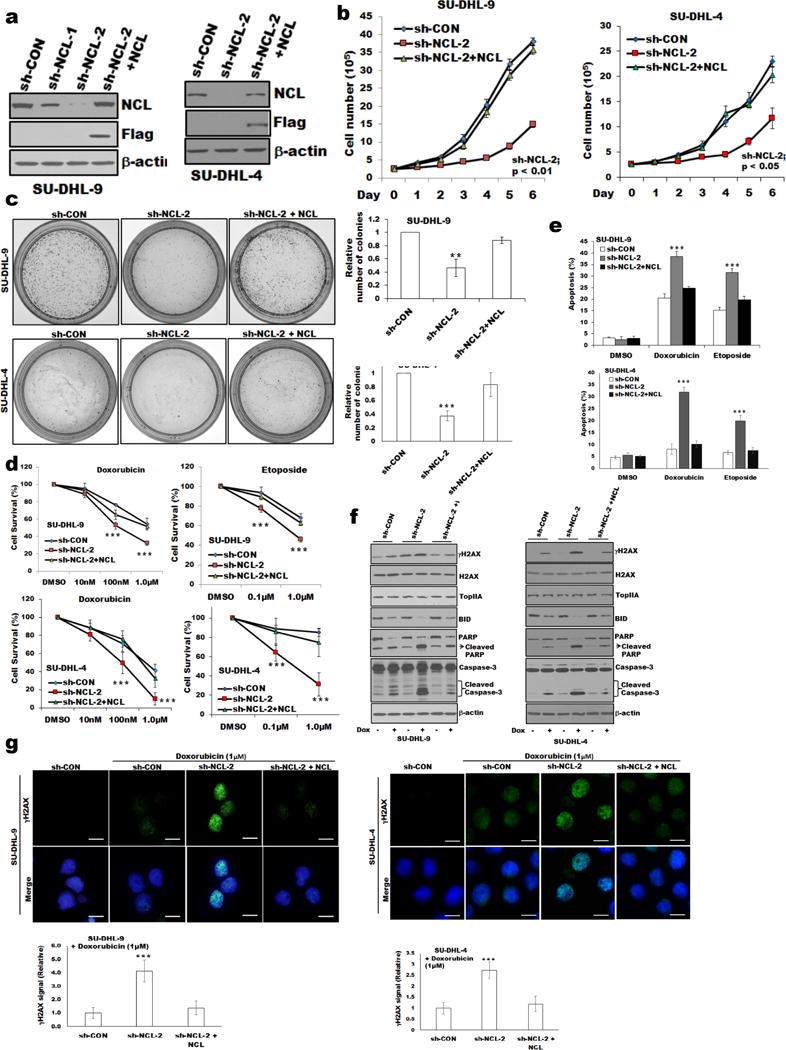
Proliferation and sensitization to doxorubicin-induced growth inhibition in nucleolin deregulated DLBCL cells.
(a) Stable knockdown of nucleolin expression in SU-DHL-4, and -9 cells using nucleolin-specific shRNAs (targeting 3′UTR). Reconstitution of nucleolin in nucleolin-knockdown cells (sh-NCL-2) was performed as described in “Methods”. Exogenous nucleolin expression in nucleolin knockdown cells was analyzed using anti-FLAG antibody. (b). Effect of nucleolin knockdown on cell proliferation. (c) Representative images of colony formation assay. The relative number of individual colonies in nucleolin-knockdown cells was significantly reduced compared to sh-control or sh-NCL-2+NCL cells, Bar=200μm. (d) MTT assay was performed as described in “Methods.” When treated with doxorubicin/etoposide, sh-NCL-2 cells showed reduced cell survival and increased percentage of apoptosis (e) compared to sh-control or sh-NCL-2+NCL cells. (f) Western blot of whole-cell lysates of SU-DHL-4, and -9 (sh-control or sh-NCL-2 or sh-NCL-2+NCL) cells treated with or without doxorubicin and analyzed for apoptotic signaling molecules, γH2AX/H2AX/TopIIA expression. (g) SU-DHL-4, and -9 cells (sh-control or sh-NCL-2 or sh-NCL-2+NCL) treated with doxorubicin were stained for γH2AX/DAPI and analyzed using fluorescence microscopy. Bar=20μm. The fluorescence intensity of γH2AX signal of 100 cells were calculated using ImageJ software and plotted. *p<0.05; **p<0.01; *** p<0.001