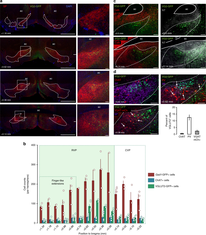
Fig. 1.
Localization and histochemical characterization of VP glutamate neurons. a Coronal images through the rostro-caudal axis of the VP of VGLUT2-GFP (VG2-GFP; green) reporter mice. b VGLUT2+ neurons were observed throughout the rostro-caudal axis but most abundant in centro-rostral VP (1791 VGLUT2+ cells, 44 sections, five mice). Gad1-GFP mice were used to assess GABA neurons (6196 Gad1-GFP+ cells, 39 sections, four mice) and all sections were stained and assessed for the cholinergic marker ChAT (1901 ChAT+ cells, 83 sections,nine mice). c Identification of the dorso-lateral subregion of the VP (VPdl) using antibodies directed against Calbindin (Calb), and the ventromedial subregion (VPvm) using antibodies directed against neurotensin (NT) show the densest cluster of VGLUT2+ cells in the VPvm. d Co-localization of VGLUT2+ cells with ChAT (1078 VGLUT2+ cells, eight VGLUT2+ ChAT+ cells, 36 sections, three mice), Parvalbumin (PV) (1692 VGLUT2+ cells, 200 VGLUT2+ PV+ cells, 44 sections, four mice), or VGAT (820 VGLUT2+ cells, 19 VGLUT2+ VGAT mCh+ cells, 16 sections, three mice). Substance P (SP) was used to delimitate VP boundaries in a and d, DAPI nuclear stain is shown in a, all distances are relative to Bregma, and cell counts or percent of VGLUT2+ cells expressed as mean ± SEM. Scale = 1 mm (a; left panels) and 100 µm (a; right panels, c and d). See also Supplementary Figure 1

