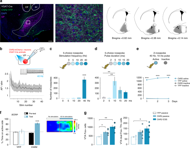
Fig. 3.
Stimulation of VP GABA neurons elicits positive reinforcement. a Expression of ChR2:YFP in the VP of VGAT-Cre mouse imaged under epifluorescent (left, scale 200 µm) or apotome (right, scale 20 µm) illumination. Counterstaining with SP (purple) and DAPI (blue), ac anterior commissure, OF optic fiber track. Spread of ChR2:YFP expression in the VP of VGAT-Cre animals (overlapped gray areas for each animal, right panel) and optic fiber tip placements (x marks the spot). b Firing of ChR2:mCh+ VP GABA neurons in response to 40-Hz photostimulation (n = 9 cells); SEM represented in gray. Inset shows representative trace; scale = 20 mV. c In a five-choice nosepoke ICSS task for VP GABA neuron stimulation mice prefer 40-Hz stimulation (n = 7). d When choosing between pulse durations, mice preferred 10 ms pulse width (n = 7). e Over 3 daily 1-h two-nosepoke ICSS sessions, mice made 1141 ± 41 nosepokes for stimulation of VP GABA neurons (n = 6 YFP controls and eight ChR2). f In an RTPP assay, mice preferred the compartment paired with stimulation of VP GABA neurons (n = 9 YFP controls and 10 ChR2). Right panel shows occupation density of an example mouse during the test trial. g Fos+ cells in the VTA increased following ICSS or passive activation of VP GABA neurons (n = 4 VGAT YFP controls, n = 5 VGAT ChR2 passive and ICSS each). The number of VTA cells double positive for the dopamine marker TH and Fos increased following ICSS for VP GABA neuron activation. *p < 0.05, **p < 0.01, ***p < 0.001. See also Supplementary Figures 5 and 6

