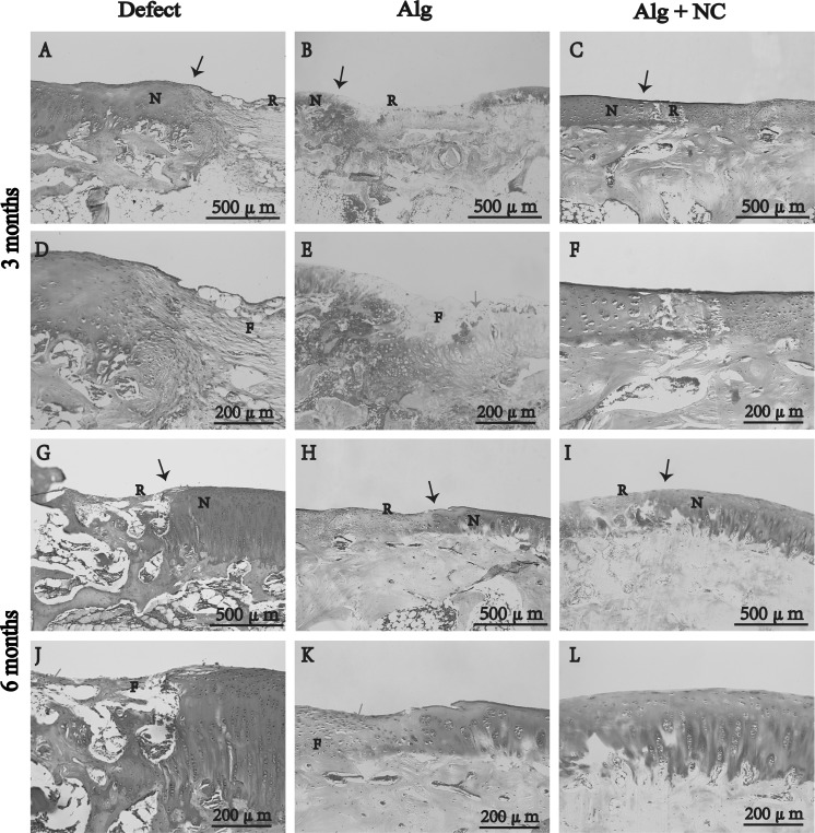
Fig. 5.
Safranin O staining of repaired tissue in three groups at 3 and 6 months after surgery. N native cartilage, R repaired tissue, F fibrous tissue. The black arrow indicate the margins of native cartilage and repaired tissue, the green arrow indicate the firoblast. d–f Are the higher magnification of repaired areas in a–c; j–l are the higher magnification of repaired areas in g–i. (Color figure online)

