Figure 2.
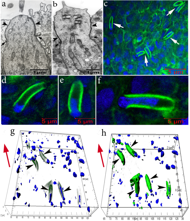
Olfactory sensory neurons in the olfactory epithelium in C. inermis. Staining for F-actin (FITC phalloidin, green) and for nuclei (DAPI, blue); transmission (a,b) and laser confocal microscopy (c-h). (a) ciliated OSN; (b) microvillous OSN; Big arrows – tight junctions; small arrows – adherens junctions; (c) OSN dendrites (marked by arrows) in the epithelium with a high content of near-membrane actin; 2D sections; (d–f) individual cells with nuclear material, which are immersed, to different degrees, in the inner space of the dendrites (2D sections); (g,h) several OSNs (marked by arrows) with nuclear material stored inside the actin case with different degrees of fluorescence filtration in the green spectrum (3D reconstructions). The red arrow indicates the direction to the surface of the olfactory epithelium.

