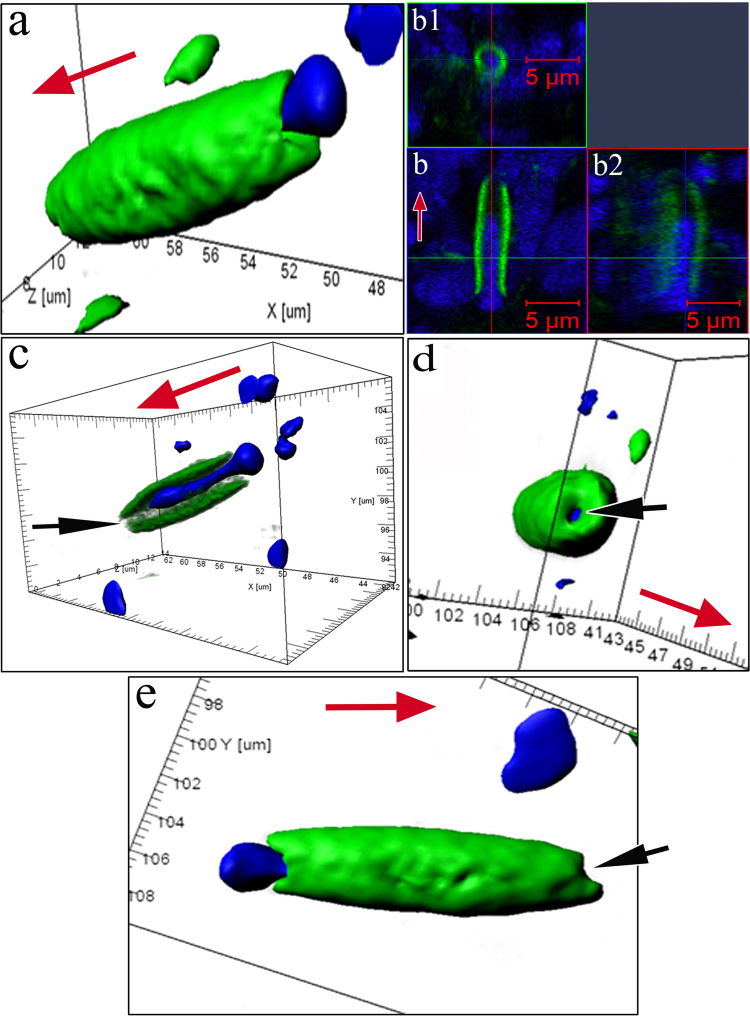
Figure 3.
Specific structural organization of OSNs during their differentiation and movement in the olfactory epithelium in C. inermis. Staining for F-actin (FITC phalloidin, green) and for nuclei (DAPI, blue); laser confocal microscopy. (A) OSN during migration (3D reconstruction); (b) A dendrite with nuclear material inside (2D section) and its orthogonal projections (b1 and b2); (c) a lengthwise cut through a iOSN; nuclear material is located inside the actin microfilaments; the front section of the dendrite is marked by an arrow; (d) apical surface of the iOSN dendrite that contains a section without actin microfilaments (marked by an arrow); (e) uneven contours of the front section of the dendrite (marked by an arrow), where the growth cone is presumably located. The red arrow indicates the direction to the surface of the olfactory epithelium.

