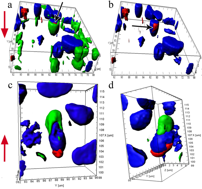
Figure 4.
Circumnuclear zones of iOSNs with growing dendrites enriched with multiple actin microfilaments in the olfactory epithelium in C. inermis; staining for F-actin (FITC phalloidin, green), for nuclei (DAPI, blue), and for mitochondria (MitoTracker®Orange CMTMRos, red); laser confocal microscopy; 3D reconstructions. (a) Mitochondria (marked by an arrow) adjacent to the nucleus in the inner space of the dendrite; (b) photo in Fig. 4a of the blue and red fluorescence channels (mitochondria marked by an arrow); (c and d) the cell body with a growing dendrite, at different angles; the upper part of the nucleus is immersed inside the dendrite; the mitochondria are located at the lower pole of the nucleus. The red arrow indicates the direction to the surface of the olfactory epithelium.

