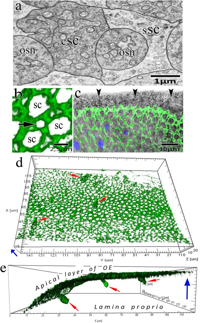
Figure 5.

The involvement of actin microfilaments in the structural organization of the olfactory epithelium and the development of receptor cells in C. inermis. (a) Transverse section through OSNs and SCs (transmission electron microscopy); (b–e) confocal microscopy; staining for F-actin (FITC phalloidin, green); (b) Mosaic organization of actin microfilaments in the olfactory epithelium (3D reconstruction); profile of the apical section of a mature OSN marked by an arrow; (c) transmitted light mode (the ciliary layer is indicated with arrows); (d) Top view of the olfactory epithelium; three OSNs (marked by arrows) incorporated in an ordered fine-mesh network of actin microfilaments in the upper layer of the olfactory epithelium (3D reconstruction); (e) the profile in Fig. 5a with the expressed actin cases of OSN dendrites (3D reconstruction); OSNs marked by arrows. Notation: OSN – olfactory sensor neuron; сSC – ciliated supporting cell; sSC – secretory supporting cell. The blue arrow shows the direction to the surface of the olfactory epithelium.
