Figure 7.
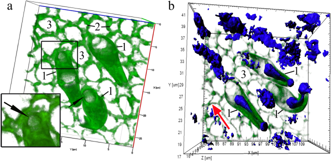
Disappearance of actin microfilaments at the tips (marked by arrows) of three OSNs whose dendrites are attached to the OE surface, in C. inermis. Staining for F-actin (FITC phalloidin, green) and for nuclei (DAPI, blue); laser confocal microscopy; 3D reconstructions. (a) А view of the OSNs from the outer side of the epithelium; the actin-lacking tip of an individual cell is marked by an arrow; the large meshes of F-actin associated with the supporting cells are clear; actin microfilaments in the axial cylinders of the dendrites are partially preserved; elongated nuclear material, the bulk of which is inside the dendrites, is visible; (b) a view of the OSNs from the lamina propria of the olfactory mucosa. Notation: 1 – OSN dendrites that contact the surface of the olfactory epithelium; 2 – Profile of the apical section of a mature OSN; 3 – Profile of the apical section of the SC. The red arrow indicates the direction to the surface of the olfactory epithelium.

