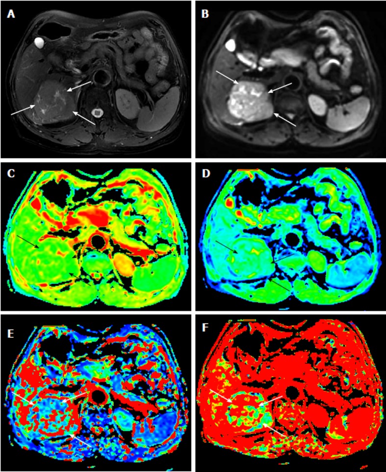
Figure 1.
Magnetic resonance images of a 65-year-old man with an 8-cm surgically verified hepatocellular carcinoma with an Edmondson-Steiner grade 1. A: T2-weighted image; B: Diffusion-weighted image with a b value of 10 s/mm2; C-F: Parametric maps (ADC, D, D*, and f, respectively) calculated from the IVIM diffusion-weighted imaging data. The tumor (white arrow) demonstrates a slightly high signal intensity on the T2-weighted image and a high signal intensity on the DWI image. The values of ADC, D, D*, and f for the ROIs of the HCC were 1.550 × 10⁻³ mm2/s, 1.110 × 10⁻³ mm2/s, 6.55 × 10⁻³ mm2/s, and 0.387, respectively, which indicated an Edmondson-Steiner grade 1 HCC. HCC: Hepatocellular carcinoma; IVIM: Intravoxel incoherent motion; DWI: Diffusion-weighted imaging.

