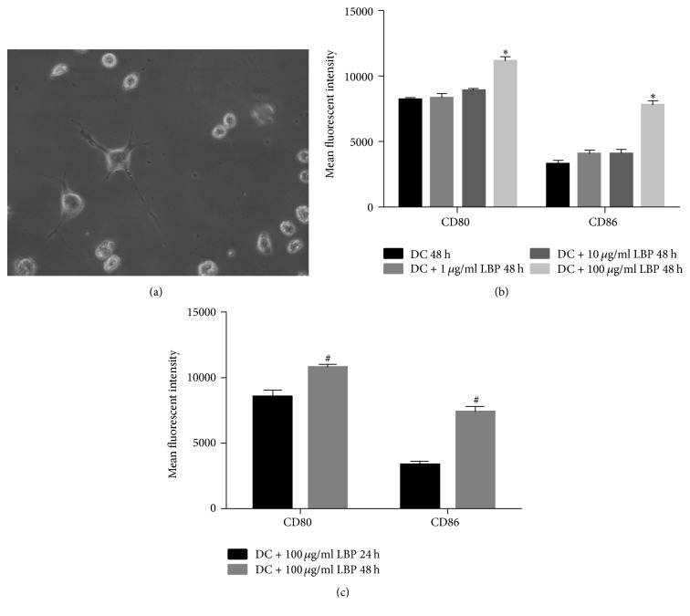
Figure 1.
Morphology and Maturation Detection of BMCs Derived DCs. (a) Seven days after induction for BMCs, 5 × 10⁵/ml DCs were incubated with LBP at 100 μg/ml for 48 h and cells morphology was detected using an inverted phase contrast microscope. The cells were characterized by long dendrites which are typical features of DCs. (b) 5 × 10⁵/ml DCs were incubated with LBP at different doses (0 μg/ml, 1 μg/ml, 10 μg/ml, and 100 μg/ml) for 48 h. The expression of CD80 and CD86 on DCs was detected using flow cytometry method. The mean fluorescent intensity of CD80 and CD86 was strongest under treatment of 100 μg/ml LBP. (c) 5 × 10⁵/ml DCs were incubated with 100 μg/ml LBP for 24 and 48 h, respectively. The expression of CD80 and CD86 on DCs was detected using flow cytometry method. The mean fluorescent intensity of CD80 and CD86 was strongest after 48 h incubation. Each assay was represented by three independent replicates. ∗P < 0.05 versus the other three groups. #P < 0.05 versus 24 h group.

