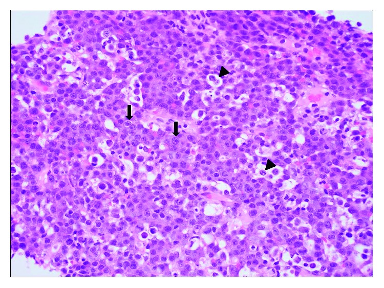
Figure 4.

High-power photomicrograph of a hematoxylin and eosin- (H/E-) stained histologic section showing effacement of the lymph node architecture by the proliferation of immunoblastic cells (arrows) with abundant amphophilic cytoplasm, eccentric round large nuclei, and prominent single macronucleoli. Scattered apoptotic bodies and tangible body macrophages (arrowheads) were seen in background (original magnification: ×400).
