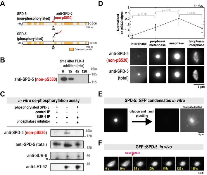
Fig. 2.
PP2ASUR-6 dephosphorylates SPD-5 and shear stresses distort and dissolve SPD-5 assemblies in vitro. (A) SPD-5 domain architecture and location of the serine 530 phospho-epitope. The non-pS530 antibody recognizes serine 530 only when not phosphorylated. (B) 200 nM of purified SPD-5 was incubated with 200 nM PLK-1+0.2 mM ATP. The reaction was analyzed at various time points by western blot using the non-pS530 antibody. (C) In vitro dephosphorylation assay. Control beads affixed to CDC-37 antibody or beads affixed to SUR-6 antibody were incubated in C. elegans embryo extract, then washed and resuspended in buffer. SPD-5 that was pre-phosphorylated in vitro by PLK-1 was then added and incubated for 90 min at 23°C and analyzed by western blot. Calyculin A was used as the phosphatase inhibitor (lane 3). The asterisk indicates a non-specific band. (D) Immunofluorescence was used to monitor relative changes in SPD-5 phosphorylation over the cell cycle. C. elegans embryos were co-stained with non-pS530 antibody and a general polyclonal SPD-5 antibody (total SPD-5). To control for changes in non-pS530 signal due to PCM growth, the ratio of the two antibody signals (non-pS530/total SPD-5) was measured for four cell cycle stages. These values were then normalized against the interphase value (mean with 95% confidence intervals; n=19 interphase, 48 prophase/metaphase, 17 anaphase, and 33 telophase centrosomes; P-values are from an unpaired t-test). (E) SPD-5 condensates were formed by incubating 500 nM SPD-5::GFP in 9% PEG-3350 for 27 min at 23°C (left), then diluted 1:10 into a 0% PEG solution and pipetted harshly (right). Dilution of SPD-5 condensates into PEG-free solution is necessary to prevent their reformation. SPD-5 condensates become more resistant to dilution as they age, thus the observed disruption and dissolution is due to pipetting (Woodruff et al., 2017; and unpublished data). The two images on the right are the same, except the contrast has been increased in the far right image to show the disrupted SPD-5 condensate. (F) Semi-permeable perm-1(RNAi) embryos expressing GFP::SPD-5 were treated with 20 µg/ml nocodazole once PCM deformation was apparent (∼250-300 s after NEBD). PCM relaxes to a spherical shape after microtubules are depolymerized (n=4 embryos).

