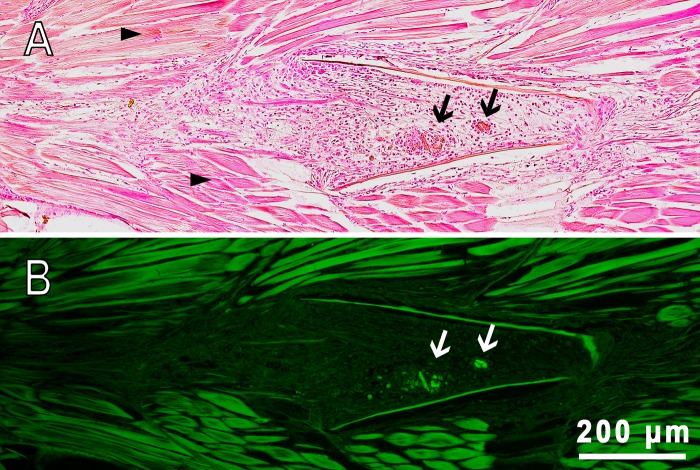
Fig. 6.
Sagittal section of fish muscles of D. rerio 22 days following the intramuscular injection of PMs-PEG. (A) Transmission image, H&E stain. (B) Fluorescence image. The arrows indicate clusters of PMs-PEG surrounded by macrophages (granulomas); arrowheads indicate normal muscle tissue composed of regular muscle fibers.

