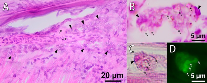
Fig. 7.
Phagocytosis of PMs-PEG in fish muscles 22 days following the intramuscular injection of PMs-PEG. (A) Microsection of D. rerio muscle, H&E stain. The arrows indicate a capillary filled with erythrocytes. The arrowheads indicate macrophages loaded with PMs-PEG (cells with granulated cytoplasm) migrating along the outer walls of blood capillaries in the lymphatic ducts. (B,C,D) Macrophages engulfing PMs-PEG. Note intact microcapsules (arrows) in the cytoplasm of phagocytes. The nuclei of the cells are marked with arrowheads. Fluorescence image (D) shows dye localization inside of intact microcapsules in the macrophage cytoplasm.

