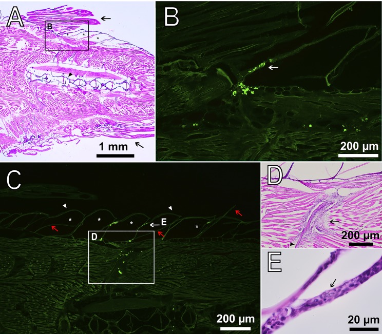
Fig. 8.
Sagittal section of D. rerio 22 days after the intramuscular injection of PMs-PEG. (A) Fish tail with dorsal (upper arrow) and anal fins (lower arrow), H&E stain. The asterisks indicate the scale pockets; the arrowhead indicates the backbone. (B) Fluorescence image scaled from A shows accumulation of macrophages loaded with PMs-PEG at the base of the dorsal fin. The arrow indicates the epidermis that covers the scales. Fluorescent images of PMs-PEG are obtained in green channel. (C) Fluorescence image of clusters of phagocytes loaded with PMs-PEG migrating from the muscle tissue toward the fish skin. The asterisks indicate the scale pockets; arrowheads indicate the epidermis; red arrows indicate the scale plates. (D) Figure scaled from C showing basophilic coloration around bone (arrowhead) indicating leucocyte infiltration and inflammation process, H&E stain. (E) Figure scaled from C shows loaded phagocyte (arrow) in the epidermis of scale, recognized by the granulated cytoplasm, H&E stain.

