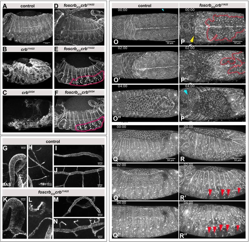
Fig. 2.
The Crb ICD is sufficient for tissue integrity of most embryonic epithelia. (A-F) Stage 12-13 embryos, stained for SAS. Dotted lines in E and F mark the disintegrated ventral epidermis. Dorsal is up, anterior left. Scale bar: 50 μm. The experiment was repeated 3 times. (G-N) Stage 12-13 foscrb; crbGX24 control (G-J) and foscrbICD crb11A22 (K-N) embryos, stained with anti-SAS. Polarity of epithelial tubes is restored in the hindgut (G,K), the Malpighian tubules (H,L), the salivary gland (I,M) and the trachea (J,N). Scale bar: 10 μm. (O-R″) Stills of time lapse movies of endogenously tagged DE-Cadherin-GFP lines. Dorsal (O-P″) and lateral (Q-R″) views of foscrb; crbGX24 control and foscrbICD crb11A22 embryos. Red dotted lines in P,P′ mark the disintegrated ventral epidermis. Yellow arrow in P, disintegrated head epidermis; cyan arrowhead in P″, recovered head epidermis; red arrowheads in R′,R″, ‘wounds’ in ventral epidermis. Anterior is to the left. Scale bar: 50 μm. The experiment was repeated 3 times.

