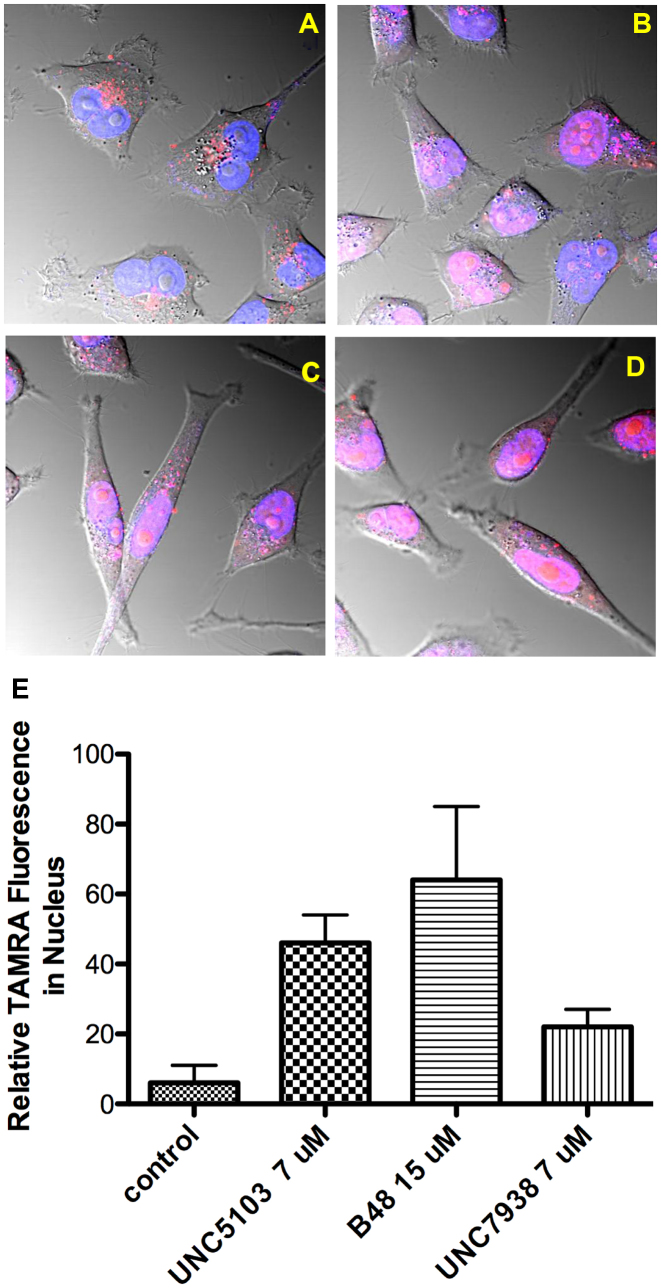
Figure 2.
OEC Effects on Re-localization of Oligonucleotide to the Nucleus. HeLa Luc 705 cells (50,000) were seeded into glass-bottom dishes and incubated at 37°C for attachment. TAMRA labeled SSO 623 (2.5 μM) was added into medium and the cells incubated for 24 h. Cells were washed with PBS and then placed back in medium and treated with OECs for 2 h or maintained as controls. Cells were rinsed after drug treatment. Live cells were imaged using a confocal microscope. (A), control cells; (B), cells treated with 15 μM B-48; (C), 7 μM UNC7938; (D), 7 μM UNC5103. Images are composites of TAMRA (red), Hoechst (blue) and DIC images and the pink coloration in indicates overlap of the TAMRA and Hoechst fluorescence. (E) Quantitates the TAMRA fluorescence in the nucleus for controls versus treated cells, as measured using Fiji software, with Hoechst stain used to delineate the nucleus. The differences between treated and control cells were significant at the 0.5% level for all compounds evaluated using the paired t-test.

