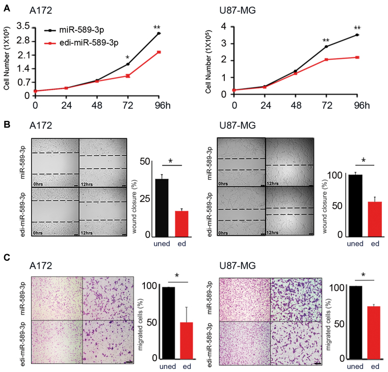
Figure 4.
Edi-miR-589–3p inhibits both proliferation and migration of glioblastoma cells. (A) A172 (2.5 × 10⁴) and U87-MG (2.5 × 10⁴) cells were transiently transfected with miR-589–3p mimic or edi-miR-589–3p mimic and cell proliferation (Trypan blue exclusion) monitored over days. Mean ± standard deviation (n = 3), *P < 0.05, **P < 0.01 (two-sided t-test). (B) 1 × 10⁵ A172 and U87-MG cells, transiently transfected with miR-589–3p (uned) or edi-miR-589–3p (ed) mimic, were seeded to confluence for scratch assay. Wound closure was monitored after 12 h (see representative pictures, scale bar 100 μm). Quantitation of wound closure is reported, mean ± standard deviation (n = 3), *P < 0.05 (two-sided t-test). (C) Transwell migration assay was performed 48 h p.t. Representative photographs of migrated cells are shown (4× and 10× magnifications). Cells were visualized by Diff Quick staining and counted 24 h post seeding. Scale bar 400 μm. Histograms show the migration ability relative to the uned-mimic (100%) (fold increase ± standard deviation, n = 3) of cells that passed through the 8 μm filter pore, *P < 0.05 (two-sided t-test).

