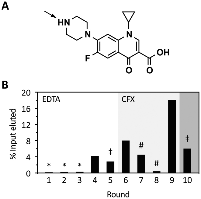
Figure 1.
Progress of in vitro selection. (A) Chemical structure of CFX. The arrow indicates the most likely attachment site to the epoxy-activated PAA-matrix. (B) Shown is the fraction of loaded RNA that could be eluted from CFX-derivatized columns after each selection round. RNA was eluted by either 20 mM EDTA (round 1–5) or 1 mM CFX (round 6–10). In the first three rounds, a negative selection was performed (*). In round 5 and 10, stringency was increased by doubling the number of column washes or a decrease in the concentration of immobilized CFX to one-tenth, respectively (‡). Pre-elution steps were performed in round seven and eight (#) (for further details see also Supplementary Table S6).

