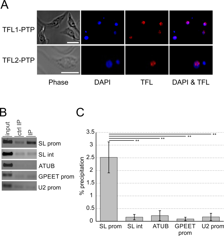
Figure 6.
TFL is localized in the nucleus and occupies the SLRNA promoter in vivo. (A) Localization of TFL1-PTP or TFL2-PTP (red) in procyclic trypanosomes. DNA was stained with DAPI (blue) showing nuclei and smaller kinetoplasts. The nucleolus can be recognized within a nucleus as a spherical structure of low DNA density. White bars in left panels represent 5 μm. (B) Anti-TFL2-PTP ChIP assay using a polyclonal anti-ProtA antibody (IP) and, in a control precipitation, a comparable nonspecific immune serum (ctrl IP). DNA from total chromatin (input) and precipitated DNA was analyzed by semi-quantitative PCR, amplifying the SLRNA promoter (SL prom), part of the SLRNA intergenic region (SL int), a fragment of the α tubulin coding region (ATUB), the GPEET procyclin promoter (GPEET prom), and the U2 snRNA gene promoter (U2 prom). (C) Corresponding qPCR analysis from three independent experiments. For each region amplified the percent precipitation was calculated and the values corrected by the corresponding values of the control precipitations. The difference in TFL2-PTP occupancy between the SLRNA promoter and the other amplified regions was statistically analyzed by paired two-tailed student's t-tests.

