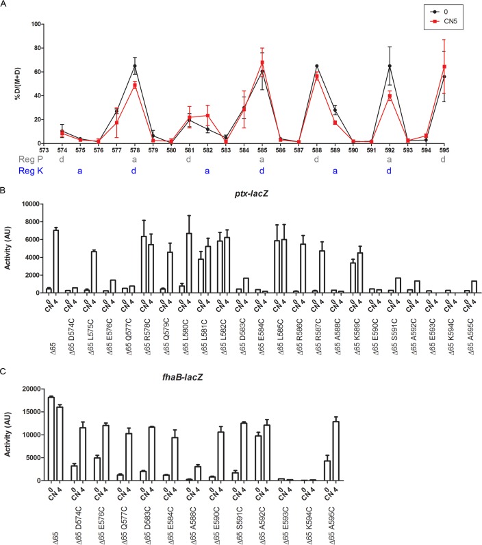
FIG 4 .
Topology and dynamics of the X linker of BvgSΔ65. (A) Cys-scanning analyses of BvgSΔ65 were performed under basal conditions (0; black curve) or after the addition of 5 mM chloronicotinate (CN 5; red curve). The proportions of dimers from two independent experiments are graphed, and the means and standard errors of the means are indicated. The “a” and “d” positions of the two coiled-coil registers (Reg P and Reg K) are indicated below amino acid positions. (B and C) The activities of the BvgS variants were determined using the ptx-lacZ (B) or fhaB-lacZ (C) reporters. The means and standard errors of the means are indicated.

