
Figure 1.
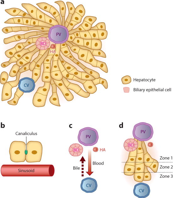
The liver lobule. (a) The liver is composed largely of two endoderm-derived cell types—hepatocytes and biliary epithelial cells—that are organized into functional units known as lobules. Blood enters the lobule through branches of the PV and HA and exits through branches of the CV to be returned to the systemic circulation. Bile makes its way to the BD, which in turn drains into the intestine to aid in digestion. (b) Blood passes through the lobule in specialized vascular channels (sinusoids). Bile is handled separately and is secreted into thin channels (canaliculi) between hepatocytes that carry the bile through the lobule and into the BD. (c) There is countercurrent flow of blood and bile: Blood flows in a portal-to-central direction, whereas bile flows in a central-to-portal direction. (d) The lobule is divided into zones, with hepatocytes closest to the PV composing zone 1 and those closest to the CV composing zone 3. Hepatocytes in different zones have different sets of functions. Abbreviations: BD, bile duct; CV, central vein; HA, hepatic artery; PV, portal vein.