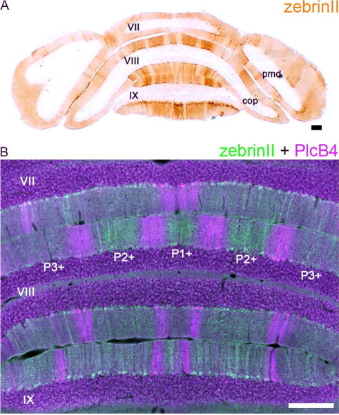
Figure 1.

(A) ZebrinII expression reveals a striking topography of parasagittal zones in the cerebellum. Shown here is a tissue section cut the posterior cerebellum of a control Swiss Webster mouse. (B) ZebrinII and PlcB4 are expressed in complementary zones (Sarna et al., 2006). Scale bars = 200 µm. Abbreviations: pmd, paramedian lobule; cop, copula pyramidis. Lobule numbers are indicated by Roman Numerals (Larsell, 1952). The zebrinII positive zones are labeled according to the standard stripe nomenclature (please see Sillitoe and Hawkes, 2002; Apps and Hawkes, 2009).
