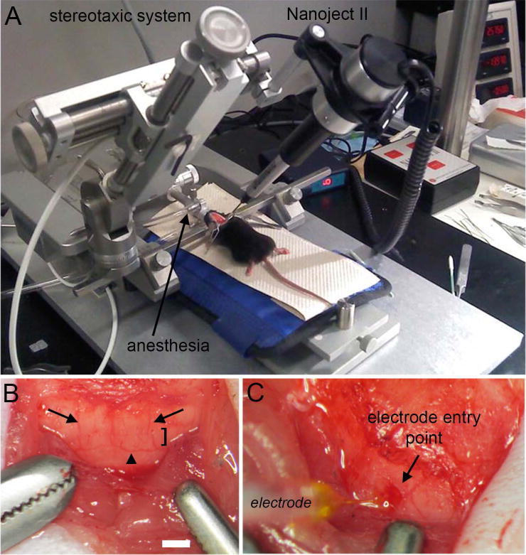
Figure 2.

(A) Surgery and injection set up for using the Nanoject II. (B) The posterior cerebellum was exposed during surgery. Lobule VIII is marked with the bracket. Lobule VIII can be identified by its unique saddle shape morphology but also by its vasculature. Dorsomedial cerebellar arteries demarcate the lateral margins of the vermis (arrows), as well as the lower visible aspect of lobule VIII that is adjacent to lobule IX (arrowhead). (C) Nanoject II mediated delivery of WGA-Alexa into lobule VIII of the cerebellum. Scale bar in B = 500 µm (= 250 µm in C).
