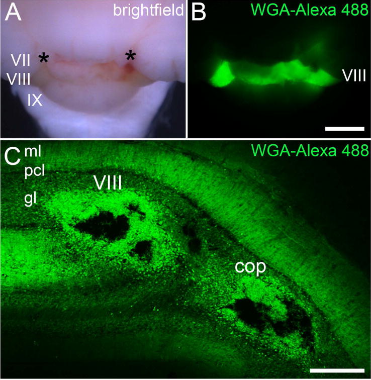
Figure 3.

(A) Brightfield image of the posterior cerebellum after injecting WGA-Alexa 488 into two separate loci (asterisks) in lobule VIII. The light green hue is due to the presence of the Fast Green that was also injected into lobule VIII. (B) Fluorescence image showing the spread of WGA-Alexa 488 within lobule VIII. (C) Coronal tissue section cut through lobule VIII. Two Nanoject II injections were made, one in the vermis portion of lobule VIII and the other in the hemispheric portion of the lobule, the copula pyramidis. Abbreviations: ml, molecular layer; pcl, Purkinje cell layer; gl, granular layer; cop, copula pyramidis. Lobule numbers are indicated by Roman Numerals (Larsell, 1952). Scale bar = 500 µm in B (applies to A) and 150 µm in C.
