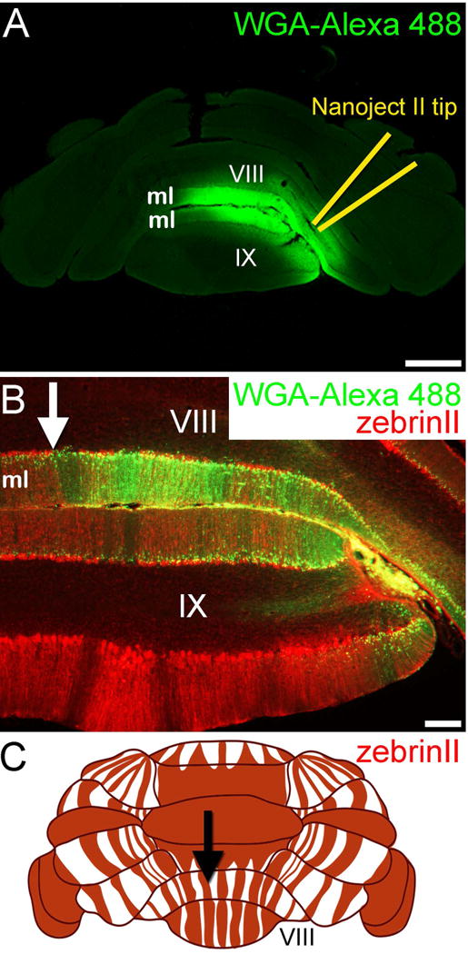
Figure 4.

(A) Nanoject injection of WGA-Alexa 488 into lobule VIII/IX results in dense tracing of the molecular layer (ml) axons. (B) The dense staining in the molecular layer is consistent with tracing of granule cell parallel fiber axons. The WGA-Alexa 488 pattern terminates at a zebrinII Purkinje cell boundary (arrow). In this experiment, the mice were given 3-acetylpyridine (3-AP) to remove climbing fiber terminals from the ml, which occurs after the drug kills inferior olive neurons. (C) Schematic of the dorsal and posterior cerebellum showing the pattern of zebrinII positive (red) and negative (white) Purkinje cell zones. The arrow is pointing to the specific zonal boundary that is also marked in panel B. Scale bar in A = 500 µm and 100 µm in B.
