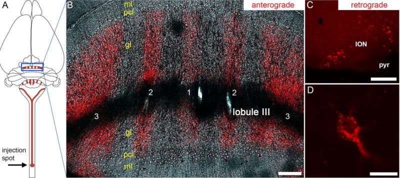
Figure 5.

(A) Schematic illustrating the strategy and overall trajectory and termination pattern of the spinocerebellar tract. The lower thoracic-upper lumbar region was targeted. The blue rectangle outlines the pattern of mossy fiber zones in the anterior cerebellum. (B) Coronal tissue section cut through the anterior cerebellum (lobule III) showing the striking pattern of mossy fiber zones after tract tracing with WGA-Alexa 555. The five major zones are labeled as 1-3, with 1 located at the cerebellar midline. (C) Injections of WGA-Alexa 555 into the spinal cord also retrogradely trace neurons, and shown here are retrogradely traced somata in the inferior olive. (D) Higher power image of a retrogradely traced inferior olive neuron showing the punctate accumulation of WGA-Alexa 555. Abbreviations: ml, molecular layer; pcl, Purkinje cell layer; gl, granular layer; ION, inferior olivary nucleus; pyr, pyramidal tract. Scale bar in B = 150 µm, scale bar in C = 200 µm, and scale bar in D = 20 µm.
