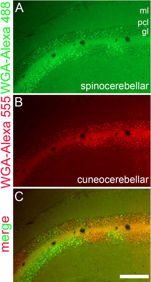
Figure 6.

Coronal tissue section cut through the anterior cerebellum showing the complementary topography of (A) spinocerebellar (WGA-Alexa 488) and (B) cuneocerebellar (WGA-Alexa 555) mossy fiber domains on one side of the cerebellum. The merged image layers are shown in panel (C). Abbreviations: ml, molecular layer; pcl, Purkinje cell layer; gl, granular layer. Scale bar = 250 µm.
