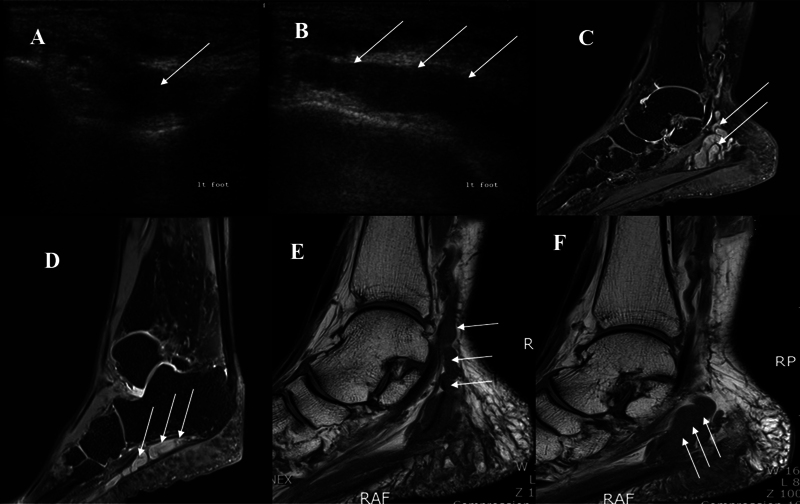
Fig. 2.

Plexiform neurofibroma. Multiple hypoechoic round masses surrounded by a hyperechoic background. The diameter of each mass measured 0.5 to 0.8 cm. ( A ) Short-axis ultrasound (US) image obtained over the calcaneus shows plexiform neurofibroma as a fusiform solid hypoechoic mass (arrow). ( B ) Long-axis US image obtained over the calcaneus shows plexiform neurofibroma as a cordlike hypoechoic mass (arrow). ( C ) T1-weighted image shows a multiloculated tubular lesion within the tarsal tunnel (arrow). ( D ) T1-weighted image shows a cordlike mass lesion below the calcaneus (arrow). ( E , F ) T2-weighted image shows a multiloculated tubulocystic lesion along the tibial nerve, lateral plantar nerve, proximal medial plantar nerve, and posterior calcaneal nerve, from the distal tibial plafond level to the fifth metatarsal base level.
