Fig. 3.

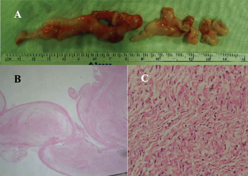
A multiloculated plexiform mass originated from the posterior tibial nerve ( A ). Plexiform arrangement (x10, hematoxylin and eosin [H&E] stain) ( B ) composed of wavy spindle cells (x400, H&E stain) ( C ) on histopathological examination.

A multiloculated plexiform mass originated from the posterior tibial nerve ( A ). Plexiform arrangement (x10, hematoxylin and eosin [H&E] stain) ( B ) composed of wavy spindle cells (x400, H&E stain) ( C ) on histopathological examination.