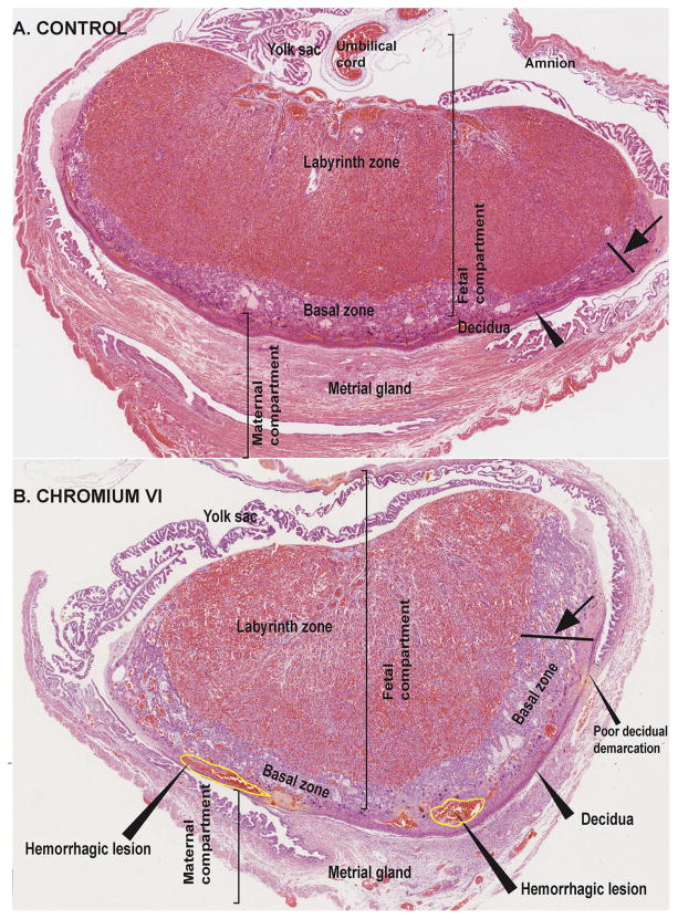
Fig. 2.
Effect of CrVI on the histopathology of whole placenta on gestation day 18.5. Pregnant mothers (n = 5) received drinking water with or without CrVI (50 ppm) from gestational day (GD) 9.5–14.5. Placentas were separated on GD 18.5 and processed for histology and sections were stained with H&E following standard protocol. H&E images of the placenta from control (A) and CrVI-treated (B) mothers, under low magnification are shown in the figure. CrVI increased hypertrophy of the basal zone (arrow); reduced the thickness of the decidual layer with poor demarcation, and caused hemorrhagic lesions above the decidual bed at the bottom layer of the basal zone, as shown in the image (arrow heads). YS – YS, LZ – labyrinth zone, BZ – basal zone and MG – MG.

